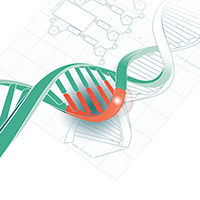
The scope and challenges of visualizing CRISPR-Cas9 gene editing in real time
Cell Gene Therapy Insights 2017; 3(1), 59-62.
10.18609/cgti.2017.009
Could you tell us about your research and how you have utilized gene editing techniques to advance your work?
Our lab is focused on human cell engineering. Specifically, we are interested in using gene editing to engineer in pathogenic mutations and gene variants into models from healthy cells, or to correct disease-associated variants within cells from patient donors. We will then use mechanistic information from these edited disease models to create new types of cell- and tissue-engineered therapies, which could be transplanted into the body.
Also, gene editing allows us to disrupt genes that we think may be important in determining cell fate. We are using editing tools to disrupt various factors inside stem cells, and then watching how those factors function during differentiation. Through these types of perturbations, we will be able to understand how to make particular cell types that are of therapeutic interest more efficiently. This can, in turn, help biomanufacturing processes.
What are some of the challenges associated with current gene editing techniques in the context of clinical translation?
I believe that controlling the heterogeneity of gene edits within a pool of human cells is one of the greatest challenges associated with current gene editing techniques. After using the CRISPR technique, typically a pool of cells will not just have one specific genotype, but a range of alleles. Even within a single cell there can be different edits on each allele. Some cells will have the desired edits; some will have undesired edits. By being better able to select only cells that have the desired edits, we ultimately hope that biomanufacturing of clinical products from human cells will be advanced.
You have developed an automated imaging platform, ArrayEdit, to determine CRISPR-Cas9’s mode of action in cells. How does visualizing gene editing in real time help to improve its application?
We are hoping that this platform will help us understand how CRISPR-Cas9 works in human cells. One advantage of using human pluripotent stem cells is standardization: we can start with one common human pluripotent stem cell line that generates a range of differentiated cell types like neurons, cardiomyocytes, myoblasts, etc. Using the identical CRISPR machinery, that is the same CRISPR nuclease and guide RNA, to edit these cells of nearly identical genotype, we aim to watch how CRISPR works in these different cell types. We hope to discover how CRISPR may be going into a particular cell differently or cutting differently. Achieving greater understanding of the molecular mechanisms is a major goal in using this platform.
Could you explain how ArrayEdit works and how this system can enhance the specificity and versatility of the CRISPR-Cas9 technique.
ArrayEdit relies on a particular surface patterned substrate, which separates out the different cell populations that are growing within a single dish, such that we can monitor them over long periods of time at high resolution through high content imaging. In the work published in Stem Cell Reports, we showed that we could easily generate the guide RNA component through in vitro transcription of synthesized oligos, at high efficiency and high throughput. That enabled us to make multiple guide RNAs easily within each well of the ArrayEdit platform, and eventually generate precise edits within human cells in each culture well.
And because our substrate is patterned, we can watch different clonal populations that may contain the desired edit(s) grow over time. One major advantage of this technique is that it multiplexes the edits easily and we can simultaneously monitor and quantify phenotypes over time through imaging, while increasing our throughput by at least an order of magnitude.
What do you see as the other potential applications and development opportunities of ArrayEdit?
I am interested in gaining additional specificity in terms of cell types that are edited. For example, in a mixed culture, which is commonly seen in stem cell-derived cultures and tissue or blood samples from patients, one would want to edit only a particular cell type, like a hematopoietic stem cell. If there is a way to image that particular cell type, and then also apply CRISPR-Cas9 in that same cell type, that would allow us to increase cell-type specificity in editing.
Another application is testing a whole range of different CRISPR assemblies. This can be used in particular to optimize materials for gene editing. We are interested in different ways to package the CRISPR machinery with biomaterials for delivery to a variety of cell/tissue types. Our work, recently published an article in Acta Biomaterialia, tested many transfection reagent mixtures with CRISPR-Cas9 with this platform.
What additional advances and new technologies do you see emerging as we try to move gene editing towards clinical application?
There is a lot of interesting work being carried out in testing different CRISPR-associated protein variants. There are high fidelity nucleases, and also entirely different proteins apart from Cas9 that people are working on, Cpf1 for instance. I have seen extensive protein engineering that I think will advance further quite quickly.
Additionally, elegant and novel delivery strategies for somatic cell editing are around the corner. Right now, emphasis is on viral delivery of the gene editing machinery. To mitigate some of the safety concerns, we are interested in non-viral delivery. There is now significant engineering R&D in both academia and industry in somatic editing for inherited diseases, partly using non-viral delivery, and I believe there will be some clinical trials coming out within the next few years.
I also foresee using patient-derived cells as a platform to test out gene editing. All of the cell types could be derived from skin biopsies and blood cells of patients through reprogramming, and then one can test a candidate gene therapy or editing technique on the patient’s own cells with their own genome on the relevant cell type. Such capabilities are extremely exciting for precision medicine, and I see the field moving in that direction.
Affiliation
Krishanu Saha
Department of Biomedical Engineering & Wisconsin Institute for Discovery, University of Wisconsin-Madison, WI, USA.

This work is licensed under a Creative Commons Attribution- NonCommercial – NoDerivatives 4.0 International License.