AAV vector and gene therapy: en route for the American dream?
Cell Gene Therapy Insights 2016; 2(5), 513-519.
10.18609/cgti.2016.066
This issue of Cell and Gene Therapy Insights puts adeno-associated vectors (AAV) on the spotlight with two thorough reviews by Dr Srivastava (University of Florida) and Dr Merten (Genethon, France). And this honor is well deserved: over the past 2 years, AAV has been propelled to the front of the line as one of the most efficient, robust, reliable and safe bio-tools to mediate long-term gene expression in human patients with genetic diseases, thanks to key advancements, both at the clinical and technical levels. Both industry and venture capitalists now capitalize on AAV’s most recent clinical successes, creating an unprecedented demand for AAV vector development and production as well as a cash flow toward AAV development and clinical use, on what was not so long ago, a purely academic research interest.
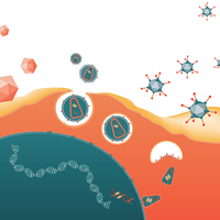
Despite this sudden momentum on AAV vectors, AAV is not the new kid on the block: AAV vectors were in fact ‘born’ more than four decades ago, and the wild-type virus, AAV serotype 2, from which the recombinant AAV are derived from, just celebrated its 50th anniversary [1,2]. In this issue of Cell & Gene Therapy Insights, Dr Srivastava provides a very elegant, yet thorough, biography of this viral vector, and we will only emphasize a few key features in this editorial: AAV is a very tiny virus, with a 26-nm diameter icosahedric protein capsid virus protecting and delivering its linear DNA genome of less than 5Kb. AAV’s eight proteins are expressed from only two major open reading frames – Rep and Cap – using a simple yet subtle ‘built-in’ approach by means of splice donor and acceptor [3] sites; AAV Rep and Cap proteins play key roles in the genome replication, gene translation, packaging, host cell binding and entry into a wide range of tissues and cell types. AAV has been described as an opportunist virus: in ‘healthy’ conditions, or in the absence of cell trauma or distress, AAV integrates its genome into a safe and latent unique site within the human genome, AAVS1; in the event of super infection by a lytic virus, such adenovirus or herpes virus, AAV is rescued from the genome, starts replicating and packaging, and quickly evades a dying host cell [4,5].
Two major events changed AAV’s journey to the human body for ever: its genome was cloned and fully sequenced in the early ‘80s [6,7], which led to the AAV first make-over in Dr Musyzca’s laboratory at the University of Florida, where it got completely degutted from its core proteins, Rep and Cap, to only retain its inverted terminal repeats (ITRs) [7,8]. Since then, AAV vectors carrying a plethora of different genetic material, from the jellyfish green fluorescent protein to the most sophisticated human genes, have been deployed to support research from basic mechanistic pathways to the treatment of genetic diseases in humans. From the original prototype to the current version of rAAV relatively little has changed in the design or concept. And it is probably necessary to re-emphasize the fact that, to date, neither the wild-type virus, nor its vector counterpart, has been linked to any human disease, making AAV the safest genetic vehicle currently developed for human gene therapy [9–11].
The list of advantages that make AAV one of the most sought after vectors for gene delivery, exhaustively described, triggered the first gene therapy trial in the mid-‘90s. However, it is only very recently that true clinical successes by means of therapeutic effect (let’s not forget that Phase I trials are to demonstrate safety, and in that sense, all Phase I trials have been successful to date) have come to light, from retinal diseases (Leber congenital amaurosis, [12,13], Leber hereditary optic neuropathy [14]) to neurological disorders (aromatic L-amino acid decarboxylase deficiency) [15], blood disorders (hemophilia B [16], lipoprotein lipase deficiency [17]) and muscular dystrophies (spinal muscular atrophy [NCT02122952] and Pompe disease [18]). With a current portfolio of over 80 clinical trials and an explosion of biotech companies, AAV is stealing the show in what most are now calling the ‘renaissance of gene therapy’. From unknown to famous, from forgotten to rediscovered, from scarce and competing government funding to multi-million dollar venture capital investment, AAV, and probably its supporters, seem to be living the American dream.
But let’s not get ahead of ourselves, AAV gene therapists. There are still major challenges on the road to clinical success, and making AAV a bio-drug accessible to patients worldwide. In his review, Dr Srivastava lists the two top challenges for AAV clinical success to be: pre-existing antibodies to AAV capsids; and tissue/cell-targeted expression. In this editorial, we want to emphasize one aspect of AAV development that is too often overlooked and underestimated: manufacturing. Full clinical and commercial success of gene therapy using AAV vectors faces the current difficulties of producing high titers, high quantities and highly pure AAV material. The current lack of well-established and validated processes for commercial scale manufacturing is not only the direct consequence of inherent technical challenges, but also of the relatively sporadic interest and funding availability dedicated to this type of research, both in academia and industry, over the past two decades.The current booming interest from industry has created an unprecedented amount of funding that benefits both industry and academia, often working in partnership, which will lead without a doubt to the manufacturing platform(s) required for the next chapter in AAV’s life: commercialization.
Dr Merten’s review in this issue provides a detailed description of AAV production methods currently in use for clinical manufacturing. The first production method was born when it was shown that the AAV genome bearing only the AAV 2 ITR could be replicated and packaged when Rep and Cap were provided in trans in the host cells, in addition to a helper virus. Since wtAAV is a defective parvovirus, some of the adenovirus or herpesvirus genes are required for its replication [7]. The first protocol relied on simultaneous transfection of two or three plasmids carrying all the required functions to produce AAV in HEK293 cells. 25 years or so later, transfection remains the most utilized method to produce rAAV, both at research and clinical grades, as recently reviewed in [19]. To date, four production methods have been implemented for the manufacture of clinical AAV products (also recently reviewed in [19,20]: transfection in both adherent or suspension cell platform [21–23]; packaging cell lines with adenovirus infection [22–24]; baculovirus expression system (BAC) [25–28]; and the herpes virus system (HSV) [29–32].
Current vector requirements to support not only clinical needs, but also extensive pre-clinicial toxicology and bio-distribution studies (a must-do to enable investigation new drug submission [IND] and approval by the American Food and drug Agency [FDA]), approach scales in the range of 1 x 10¹⁵ to 1 x 10¹⁶ AAV vector genomes (vg). It is already foreseeable that these needs will soon commonly surpass 1 x 10¹⁷ while commercial-scale requirements will exceed capacity of 1 x 10¹⁸ vg. In other words, and to quote Dr Srivastava’s more emphatic terminology, we are looking at producing quadrillions (1 x 10¹⁵) to quintillions (1 x 10¹⁸) of AAV in the next 2 years.
Yields between 1 x 10¹⁵ to 1 x 10¹⁶ are currently achievable in a limited number of laboratories, often benefiting from high capacity in terms of equipment, space and labor, combined with processes that remain to be tailored for commercial scale. The first commercially produced AAV was Glybera® (alipogene tiparvovec, UniQure), an AAV1-based drug for the treatment of lipoprotein lipase deficiency in adults [17,33]. To date, no AAV drugs have been approved nor submitted for approval in the USA, and it is not known which of the four methods mentioned above will support the first market approval of an AAV drug. The HSV packaging cell lines and transfection have been FDA approved and used in clinical trials in the USA. The capacity in terms of yields and the safety profiles may be giving the HSV system a slight lead over its other counterparts, yet several large biotech companies have invested in the BAC system to support their large-scale manufacturing and future market-scale production. It will take several years for the scientific community to gather sufficient data, both from pre-clinical and clinical studies, as well as from technical reports during the implementation phases of their method of choice, and to determine the feasibility, time constraints and cost of such implementation. As of today, it is foreseeable that several methods will be implemented and used, based not only on data-driven decisions but also, as we will discuss below, intellectual property and legal rights to such methods. As a result, the community, together with the FDA will need to establish clear standards to support bio-equivalency studies and demonstrate bio-compatibility across the different methods used.
Of the four main production methods, all have benefits and disadvantages. There is an overwhelming agreement that the transfection method in adherent format (HEK293 or HEK293T grown in multi-layer flasks) will not support large clinical and marketing scale production, since scalability is directly proportional to the surface area (i.e., the number of flasks) needed. A typical GMP manufacturing campaign requires more than 100 10-layer cellstacks for yields in the 1 x 10¹⁵ vg of clinical product [19,34], and generating more than 1 x 10¹⁶ vg would require greater than 500–1000 cell factories. Although technically possible, it is likely not a viable option for most if not all manufacturing facilities with regard to manpower, space and cost limitations. The most appealing methods rely on suspension culture, whether using chemical (transfection) or biological reagents such BAC or HSV. Based on published yields, the two methods that seem the most comparable are BAC and tHSV with yields often more than 105 vg/cell of purified material [25,31; Adamson et al., manuscript in preparation]. Both methods require relatively extensive upstream work with reagent development, either recombinant BAC or HSV to support AAV production, and include production and release testing of viral banks (raw material) or virus-infected cell banks. However, transfection still relies on a massive amount of plasmid to be produced at GMP-grade, and all things considered, could result in a higher cost based on lower yield per cell. Methods relying on biological raw materials like BAC or HSV also face challenges with regards to the viral stock genetic and infectious stability, as well as extensive safety testing to determine the amounts of residual viral materials (DNA, protein, wild-type or replication competent viral particles; recently reviewed in [19], and in this issue). Yet, as discussed above, the data currently available is not sufficient to determine the true technical challenges and cost of each method.
Another daunting aspect in the field is the complex yet unavoidable difficulty to consistently determine biological parameters of clinical AAV drugs based on the diversity and variability of quality control assays available [19,35]. Cross-referencing INDs would be the easiest, fastest and cheapest way to support clinical research of similar AAV products. Yet the inherent assay-to-assay or lab-to-lab variability makes it almost impossible to compare clinical doses from one trial to another, unless biological conversion rates are established between the different laboratories. Furthermore, with each new method, assays need to be developed to determine potential process-residual or impurities. Guidance on the level of impurities acceptable for increased human doses, especially in the context of systemic administrations, is still uncharted territory. Last but not least, stability of the purified particles may vary according to the methods used for production, purification and final formulation buffer, which will require formal stability studies to be initiated over several years.
Recently, a new type of challenge, neither biological, technical nor clinical, has plagued the scientific community and impacted decisions and research pace: intellectual property. Legal rights to use AAV reagents, sometimes created decades ago, have become a major roadblock to most academic and industry institutions, and often resulted in significant time, energy and cost investment. In many instances, it seems easier to re-invent the wheel than negotiate the rights to use the one available.
In conclusion, long-term clinical success of AAV is interweaved with making sure that the demand will be honored by implementing the necessary methods to produce large amounts of high-quality and high-potency AAV particles. Manufacturing capability not only requires the protocols in place, funding, legal rights, but also highly trained experts in both the academic and industry settings and access to manufacturing suites. Continued and substantive efforts to invest in research, training, develop robust and universal quality assays, increase the availability of manufacturing centers, both academic and industry-driven, streamline legal negotiations, and FDA approval, will pave AAV’s road to success.
Enjoy reading this exciting issue of Cell & Gene Therapy Insights!
financial & competing interests disclosure
The author has no relevant financial involvement with an organization or entity with a financial interest in or financial conflict with the subject matter or materials discussed in the manuscript. This includes employment, consultancies, honoraria, stock options or ownership, expert testimony, grants or patents received or pending, or royalties. No writing assistance was utilized in the production of this manuscript.
References
1.Hastie E, Samulski RJ. Adeno-associated virus at 50: a golden anniversary of discovery, research, and gene therapy success – a personal perspective. Hum. Gene Ther. 2015; 26(5), 257–65. CrossRef
2.Muzyczka N, Berns KI. AAV’s Golden Jubilee. Mol. Ther. 2015; 23(5): 807–8. CrossRef
3.Sonntag F, Schmidt K, Kleinschmidt JA. A viral assembly factor promotes AAV2 capsid formation in the nucleolus. Proc. Natl Acad. Sci. USA 2010; 107(22): 10220–5. CrossRef
4.Berns KI, Giraud C. Biology of adeno-associated virus. Curr. Top. Microbiol. Immunol. 1996; 218, 1–23. CrossRef
5.Berns KI, Giraud C. Adenovirus and adeno-associated virus as vectors for gene therapy. Ann. NY Acad. Sci. 1995; 772, 95–104. CrossRef
6.Samulski RJ, Berns KI, Tan M, Muzyczka N. Cloning of adeno-associated virus into pBR322, rescue of intact virus from the recombinant plasmid in human cells. Proc. Natl Acad. Sci. USA 1982; 79(6), 2077–81. CrossRef
7.Samulski RJ, Srivastava A, Berns KI, Muzyczka N. Rescue of adeno-associated virus from recombinant plasmids, gene correction within the terminal repeats of AAV. Cell 1983; 33(1), 135–43. CrossRef
8.Hermonat PL. The first adeno-associated virus gene transfer experiment, 1983. Hum. Gene Ther. 2014; 25(6), 486–7. CrossRef
9.Flotte TR, Carter BJ. Adeno-associated virus vectors for gene therapy. Gene Ther. 1995; 2(6), 357–62. Reference
10.Daya S, Berns KI. Gene therapy using adeno-associated virus vectors. Clin. Microbiol. Rev. 2008; 21(4), 583–93. CrossRef
11.Kumar SR, Markusic DM, Biswas M, High KA, Herzog RW. Clinical development of gene therapy: results and lessons from recent successes. Mol. Ther. Methods Clin. Dev. 2016; 3, 16034. CrossRef
12.Jacobson SG, Cideciyan AV, Ratnakaram R et al. Gene therapy for leber congenital amaurosis caused by RPE65 mutations: safety and efficacy in 15 children and adults followed up to 3 years. Arch. Ophthalmol. 2012; 130(1), 9–24. CrossRef
13.Testa F, Maguire AM, Rossi S et al. Three-year follow-up after unilateral subretinal delivery of adeno-associated virus in patients with Leber congenital Amaurosis type 2. Ophthalmology 2013; 120(6), 1283–91. CrossRef
14.Koilkonda RD, Yu H, Chou TH et al. Safety and effects of the vector for the Leber hereditary optic neuropathy gene therapy clinical trial. JAMA Ophthalmol. 2014; 132(4), 409–20. CrossRef
15.Hwu WL, Muramatsu S, Tseng SH et al. Gene therapy for aromatic L-amino acid decarboxylase deficiency. Sci. Transl. Med. 2012; 4(134), 134ra61. CrossRef
16.Nathwani AC, Reiss UM, Tuddenham EG et al. Long-term safety and efficacy of factor IX gene therapy in hemophilia B. N. Engl. J. Med. 2014; 371(21), 1994–2004. CrossRef
17.Gaudet D, Stroes ES, Méthot J et al. Long-Term Retrospective Analysis of Gene Therapy with Alipogene Tiparvovec and Its Effect on Lipoprotein Lipase Deficiency-Induced Pancreatitis. Hum. Gene Ther. 2016; Epub ahead of print. CrossRef
18.Byrne PI, Collins S, Mah CC et al. Phase I/II trial of diaphragm delivery of recombinant adeno-associated virus acid alpha-glucosidase (rAAaV1-CMV-GAA) gene vector in patients with Pompe disease. Hum. Gene Ther. Clin. Dev. 2014; 25(3), 134–63. CrossRef
19.Clement N, Grieger JC. Manufacturing of recombinant adeno-associated viral vectors for clinical trials. Mol. Ther. Methods Clin. Dev. 2016; 3, 16002. CrossRef
20.Wright JF. New adeno-associated virus strategies to support momentum in the clinic. Hum. Gene Ther. 2011; 22(5), 519–21.
CrossRef
21.Grieger JC, Soltys SM, Samulski RJ. Production of Recombinant Adeno-associated Virus Vectors Using Suspension HEK293 Cells and Continuous Harvest of Vector From the Culture Media for GMP FIX and FLT1 Clinical Vector. Mol. Ther. 2016; 24(2), 287–97. CrossRef
22.Clark KR, Voulgaropoulou F, Fraley DM, Johnson PR. Cell lines for the production of recombinant adeno-associated virus. Hum. Gene Ther. 1995; 6(10), 1329–41. CrossRef
23.Liu XL, Clark KR, Johnson PR. Production of recombinant adeno-associated virus vectors using a packaging cell line and a hybrid recombinant adenovirus. Gene Ther. 1999; 6(2), 293–9. CrossRef
24.Martin J, Frederick A, Luo Y et al. Generation and characterization of adeno-associated virus producer cell lines for research and preclinical vector production. Hum. Gene Ther. Methods 2013; 24(4), 253–69. CrossRef
25.Cecchini S, Virag T, Kotin RM. Reproducible high yields of recombinant adeno-associated virus produced using invertebrate cells in 0.02- to 200-liter cultures. Hum. Gene Ther. 2011; 22(8), 1021–30. CrossRef
26.Bryant LM, Christopher DM, Giles AR et al. Lessons learned from the clinical development and market authorization of Glybera. Hum. Gene Ther. Clin. Dev. 2013; 24(2), 55–64. https://doi.org/10.1089/humc.2013.087″ target=”_blank”>CrossRef
27.Kotin RM. Large-scale recombinant adeno-associated virus production. Hum. Mol. Genet. 2011; 20(R1), R–6. CrossRef
28.Felberbaum RS. The baculovirus expression vector system: a commercial manufacturing platform for viral vaccines and gene therapy vectors. Biotechnol. J. 2015; 10(5), 702–14. CrossRef
29.Chulay JD. Preclinical evaluation of a recombinant adeno-associated virus vector expressing human alpha-1 antitrypsin made using a recombinant herpes simplex virus production method. Hum. Gene Ther. 2011; 22(2), 155–65. CrossRef
30.Thomas DL, Wang L, Niamke J et al. Scalable recombinant adeno-associated virus production using recombinant herpes simplex virus type 1 coinfection of suspension-adapted mammalian cells. Hum. Gene Ther. 2009; 20(8), 861–70. CrossRef
31.Adamson-Small L. A scalable method for the production of high-titer and high-quality adeno-associated type 9 vectors using the HSV platform. Mol. Ther. Methods Clin. Dev. 2016; 3, 16031. CrossRef
32.Clement N, Knop DR, Byrne BJ. Large-scale adeno-associated viral vector production using a herpesvirus-based system enables manufacturing for clinical studies. Hum. Gene Ther. 2009; 20(8), 796–806. CrossRef
33.UniQure: Website
34.Allay JA. Good manufacturing practice production of self-complementary serotype 8 adeno-associated viral vector for a hemophilia B clinical trial. Hum. Gene Ther. 2011; 22(5): 595–604. CrossRef
35.Wright JF, Zelenaia O. Vector characterization methods for quality control testing of recombinant adeno-associated viruses. Methods Mol. Biol. 2011; 737, 247–78. CrossRef
Affiliations
Nathalie Clément
Powell Gene Therapy Center, Department of Pediatrics, University of Florida, 1200 Newell Drive, Gainesville, FL, USA

This work is licensed under a Creative Commons Attribution- NonCommercial – NoDerivatives 4.0 International License.